کاروان سلامتی
کاروان سلامتی در همه موارد......کاروان سلامتی
کاروان سلامتی در همه موارد......Achilles tendon
Achilles tendon
From Wikipedia, the free encyclopedia
| Achilles tendon | |
|---|---|
 | |
| Posterior view of the foot and leg, showing the Achilles tendon (tendo calcaneus). The gastrocnemius muscle is cut to expose the soleus. | |
 | |
| Lateral view of the human ankle, including the Achilles tendon | |
| Latin | tendo calcaneus, tendo Achillis |
| Gray's | subject #129 483 |
| MeSH | Achilles+tendon |
| Dorlands/Elsevier | t_04/12793915 |
- This is about vertebrate anatomy. For the metaphor of a single vulnerable spot, see Achilles' heel.
The Achilles tendon (or occasionally Achilles’ tendon) also known as the calcaneal tendon or the tendocalcaneous is a tendon of the posterior leg. It serves to attach the gastrocnemius (calf) and soleus muscles to the calcaneus (heel) bone.
Contents[hide] |
[edit] Anatomy
The Achilles tendon is the tendonous extension of three muscles in the lower leg: gastrocnemius, soleus, and plantaris. In humans, the tendon passes behind the ankle. It is the thickest and strongest tendon in the body. It is about 15 cm long, and begins near the middle of the leg, but receives fleshy fibers on its anterior surface, almost to its lower end. Gradually becoming contracted below, it is inserted into the middle part of the posterior surface of the calcaneus, a bursa being interposed between the tendon and the upper part of this surface. The tendon spreads out somewhat at its lower end, so that its narrowest part is about 4 cm. above its insertion. It is covered by the fascia and the integument, and stands out prominently behind the bone; the gap is filled up with areolar and adipose tissue. Along its lateral side, but superficial to it, is the small saphenous vein. The achilles' muscle reflex tests the integrity of the S1 spinal root.
[edit] Nomenclature
The oldest-known written record of the tendon being named for Achilles is in 1693 by the Flemish/Dutch anatomist Philip Verheyen. In his widely used text Corporis Humani Anatomia, Chapter XV, page 328, he described the tendon's location and said that it was commonly called "the cord of Achilles" ("quae vulgo dicitur chorda Achillis").
The name Achilles' heel comes from Greek mythology. His mother, the goddess Thetis, received a prophecy of her son's death. In order to protect him, she dipped him into the River Styx, which protected his entire body from harm. However, in order to dip him into the river, she needed to grab onto his heel. During the Trojan War, Achilles was struck on his unprotected heel by a poisoned arrow, which killed him.
Because eponyms have no relationship to the subject matter anatomical eponyms are being replaced by descriptive terms. The current terminology for Achilles tendon is calcaneal tendon.
[edit] Role in disease
Achilles tendinitis is inflammation of the tendon, generally due to overuse of the affected limb or as part of a strain injury. More common is Achilles tendinosis, a degenerative condition with inflammation of the tendon, often accompanied by pain and swelling of the surrounding tissue and paratendon. Maffulli et al. suggested that the clinical label of tendinopathy should be given to the combination of tendon pain, swelling and impaired performance. Achilles tendon rupture is a partial or complete break in the tendon; it requires immobilisation or surgery. Xanthoma can develop in the Achilles tendon in patients with familial hypercholesterolemia.
[edit] Treatment of Achilles tendon damage
Initial treatment of damage to the tendon is generally nonoperative. Orthotics can produce early relief to the tendon by the correction of malalignments, non-steroidal anti-inflammatory drugs (NSAIDs) are generally to be avoided as they make the more-common tendinopathy (degenerative) injuries worse[citation needed]; though they may very occasionally be indicated for the rarer tendinitis (inflammatory) injuries. Physiotherapy by eccentric calf stretching under resistance is commonly recommended, usually in conjunction with podiatric insoles or heel cushioning. According to reports by Hakan Alfredson, M.D., and associates of clinical trials in Sweden, the pain in Achilles tendinopathy arises from the nerves associated with neovascularization and can be effectively treated with 1-4 small injections of a sclerosant. In a cross-over trial, 19 of 20 of his patients were successfully treated with this sclerotherapy.
Where tendon rupture is concerned, there are three main types of treatment: the open and the percutaneous operative methods, and nonoperative approaches.
[edit] Additional images
|
The popliteal, posterior tibial, and peroneal arteries. |
Back of left lower extremity. |
 The mucous sheaths of the tendons around the ankle. Lateral aspect. |
The mucous sheaths of the tendons around the ankle. Medial aspect. |

پارگی تاندون آشیل ( چهارشنبه 8 شهریور 1385 )
پیش زمینه
پارگی تاندون آشیل بطور معمول در افراد جوان و فعال و سالم 30 الی 50 ساله که هیچ سابقه ای از درد مچ یا پا نداشته اند رخ می دهد. اغلب پارگی های تاندون آشیل 2-1 اینچ بالاتر از محل اتصال آن به استخوان پاشنه پا ایجاد می شود و فقط در تنیسورها است که امکان پارگی زرد پی آشیل در محل اتصال آن به عضله پشت ساق زیاد است.
عدم آمادگی کامل بدنی, سن بالا و کشش بیش از حد اعمال کردن بر روی عضله پشت ساق از علل مهم خطر برای ایجاد پارگی تاندون آشیل می باشند اما شایعترین رخدادی که سبب پارگی آشیل می گردد اعمال نیروی کششی بر روی پایی است که در محل مچ به سمت عقب خم شده است. مثل شروع استارت دو های سرعت 100 متر. البته پارگی و آسیب زردپی آشیل می تواند در اثر اعمال ضربه مستقیم به آن نی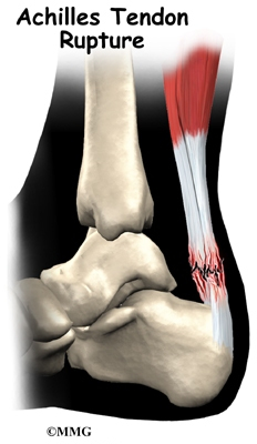 ز رخ دهد.
فرکانس
میزان شیوع پارگی تاندون آشیل مشخص نیست اگر چه در مردان شایعتر از زنان است و در دهه های 3 تا 5 زندگی هم شیوع بیشتری دارد.
آناتومی کاربردی
زردپی آشیل بزرگترین و قوی ترین تاندون بدن انسان است. طول این تاندون در حدود 15 سانتی متر است و ادامه تاندونی عضله سه سر پشت ساق محسوب می گردد. تاندون آشیل باید زاویه حدود 30 درجه به استخوان پاشنه پا اتصال می یابد. خونرسانی آن از انتهای عضلانی آن و نیز از عروقی که از استخوان پاشنه به آن وارد می شوند تامین می گردد. 6-2 سانتی متری انتهای تحتانی آن عروق خونی ندارد و از قسمت محیطی و به طریقه انتشار تغذیه می شود و همین بخش بدون عروق است که در تمرینهای مداوم و فشارهای مختلف به سبب کمبود خونرسانی می تواند دچار آسیب دیدگی و پارگی شود.
بیومکانیک ویژه ورزش
عمل عضله سه سر پشت ساق که تاندون آشیل آنرا به استخوان پاشنه پا اتصال می دهد خم کردن زانو و خم کردن به عقب مچ پا ( پلانتار فلکسیون ) است. خم شدن به عقب مچ پا در حرکات ورزشی اهمیت زیادی دارد مثل دویدن, پریدن و ایستادن و بالا رفتن از جایی مثل کوهنوردی . در خلال دویدن فشارهای معادل 10 برابر وزن بدن بر روی تاندون آشیل وارد می آید که اگر آسیب قبلی در آن وجود داشته باشد مستعد آسیب مجدد خواهد بود.
شرح حال
• فرد آسیب دیده معمولاً بیان می کند که احساس پاره شدن چیزی در پشت پا داشته و بدنبال آن دچار درد شدیدی شده است.
• شاید بیان کند که در اثر اصابت ضربه و یا بازشدن ناگهانی پا در وضعیتی که مچ پا به عقب خم بوده این درد ایجاد شده است.
• امکان دارد که قبل از این درد شدید قبلاً در هنگام ورزش دچار درد خفیف پشت مچ پا می شده که اگر این امر را بیان کند نشانه آسیب قبلی یا زمینه ای تاندون آشیل مثل التهاب آن وجود دارد. که در زمینه این آسیب قبلی حال دچار پارگی تاندون شده است.
• سابقه ای از تزریق داخل تاندونی استروئیدها که می تواند سبب ضعف تاندون شود هم باید پرسیده شود.
• فرد بیان می کند که نمی تواند روی انگشتان پا بایستد یا از روی زمین بلند شود یا اینکه بدود.
• باید پرسیده شود که آیا قبلاً هم در همین پا دچار پارگی شده است یا خیر.
معاینه
• باید تمام طول عضله سه سر پشت ساق و تاندون آشیل معاینه و لمس شود و به هر گونه تورم حساسیت در لمس و نقص و فقدان تاندونی توجه گردد.
• فرد در خم کردن مچ پا به عقب در برابر مقاومت دست ما ناتوان است.
• انجام تستهای تخصصی مثل : Hyper dorsiflexion و Thompson test و O’Brien needle test .
علل پارگی تاندون آشیل
ریسک فاکتورهای پارگی تاندون آشیل عبارتند از :
1- ورزش کردن ناپیوسته و گاهگاهی
2- سن بین 50-30 سال
3- پارگی یا آسیب قبلی تاندون آشیل
4- تزریق قبلی استروئید داخل تاندون
5- تغییر شدید در میزان تمرین یا شدت تمرین.
6- شرکت در فعالیتهای جدید که در آنها مهارت کافی نداریم.
بررسی های تصویری
• رادیوگرافی مچ پا می تواند تورم بافت نرم , کلسیفیکاسیون و کندگی همزمان بخشی از استخوان پاشنه پا را نشان دهد. رادیوگرافی برای رد کردن اختلالات استخوانی و شکستگی های همراه بسیار مفید است.
• MRI و سونوگرافی برای تایید تشخیص کمک می کنند اما اهمیت این روش در پارگی های جزئی تاندون آشیل است که در رادیوگرافی دیده نمی شود.
• سونوگرافی ساده, سریع, ارزان و قابل تکرار است و با معاینه دینامیک می توان ضخامت تاندون و میزان فاصله خالی در اثر پاره شدن را تشخیص داد. اما بشدت وابسته به شخصی است که آنرا انجام می دهید.
• MRI برای تشخیص پارگی های جزئی تاندون بسیار حساس است. همچنین برای تغییرات تخریبی مزمن تاندون, التهاب غلاف تاندونی, پارگی کامل آشیل و ضخیم شدگی تاندون تشخیصی است. اما بررسی دینامیک با آن امکانپذیر نبوده و نیز گران است.
درمان
در صورت پارگی کامل نیاز به انجام جراحی وجود دارد. پس از بهبودی کافی تاندون با تشخیص پزشک فیزیوتراپی باید صورت گیرد تا قدرت عضلانی و استحکام تاندونی و حس عمقی مناسب از مفصل شکل گیرد.
I think all black toenail abnormalities,ridging,discoloration or splitting can be symptomatic of numerous underlying problems ranging from malnutrition to heart disease.Please have a health care specialist look at your nails to rule out any threat to your general health.Good luck.